
Fri, Nov 20, 2026
[Archive]
Volume 11, Issue 4 (11-2026)
J Nutr Food Secur. 2026, 11(4): 683-696 |
Back to browse issues page


Download citation:
BibTeX | RIS | EndNote | Medlars | ProCite | Reference Manager | RefWorks
Send citation to:


BibTeX | RIS | EndNote | Medlars | ProCite | Reference Manager | RefWorks
Send citation to:
Raoofi A, Shafaei B, Ataee Disfani R, Mozaffari-Khosravi H, Sangouni A A. Non-Alcoholic Fatty Liver Disease, Gut Microbiota and Synbiotics: A Review of the Experimental and Clinical Evidence. , J Nutr Food Secur.. 2026; 11 (4) :683-696
URL: http://jnfs.ssu.ac.ir/article-1-1255-en.html
URL: http://jnfs.ssu.ac.ir/article-1-1255-en.html
Amir Raoofi  , Behnam Shafaei , Reza Ataee Disfani , Hassan Mozaffari-Khosravi , Abbas Ali Sangouni *
Health Research Center, Life Style Institute, Baqiyatallah University of Medical Sciences, Tehran, Iran
Full-Text [PDF 973 kb]
(137 Downloads)
| Abstract (HTML) (1777 Views)
1 Cellular and Molecular Research Center, Department of Anatomy, Sabzevar University of Medical Sciences, Sabzevar, Iran.2 Children Growth Disorder Research Center, Shahid Sadoughi University of Medical Sciences, Yazd, Iran; 3 Student Research Committee, Sabzevar University of Medical Sciences, Sabzevar, Iran; 4 Department of Nutrition, School of Public Health, Shahid Sadoughi University of Medical Sciences, Yazd, Iran; 5 Health Research Center, Life Style Institute, Baqiyatallah University of Medical Sciences, Tehran, Iran; 6 Department of Nutrition and Food Hygiene, Faculty of Health, Baqiyatallah University of Medical Sciences, Tehran, Iran.
Full-Text: (51 Views)
Non-Alcoholic Fatty Liver Disease, Gut Microbiota and Synbiotics:
A Review of the Experimental and Clinical Evidence
Amir Raoofi; PhD 1, Behnam Shafaei; MD 2, Reza Ataee Disfani; MD 3, Hassan Mozaffari-Khosravi; PhD 4 & Abbas Ali Sangouni; PhD *5,6
A Review of the Experimental and Clinical Evidence
Amir Raoofi; PhD 1, Behnam Shafaei; MD 2, Reza Ataee Disfani; MD 3, Hassan Mozaffari-Khosravi; PhD 4 & Abbas Ali Sangouni; PhD *5,6
1 Cellular and Molecular Research Center, Department of Anatomy, Sabzevar University of Medical Sciences, Sabzevar, Iran.2 Children Growth Disorder Research Center, Shahid Sadoughi University of Medical Sciences, Yazd, Iran; 3 Student Research Committee, Sabzevar University of Medical Sciences, Sabzevar, Iran; 4 Department of Nutrition, School of Public Health, Shahid Sadoughi University of Medical Sciences, Yazd, Iran; 5 Health Research Center, Life Style Institute, Baqiyatallah University of Medical Sciences, Tehran, Iran; 6 Department of Nutrition and Food Hygiene, Faculty of Health, Baqiyatallah University of Medical Sciences, Tehran, Iran.
| ARTICLE INFO | ABSTRACT | |
| REVIEW ARTICLE | Background: Non-alcoholic fatty liver disease (NAFLD) is a multi-cause disease. Several factors are involved in the pathogenesis of NAFLD. Recently, the role of gut microbiota in the pathogenesis of NAFLD has attracted the attention of researchers. This study was performed to review the role of gut microbiota in the pathogenesis of NAFLD. In addition, the mechanisms and clinical effects of synbiotics on NAFLD were reviewed. Methods: A narrative review of the most recent advances was conducted regarding the role of dysbiosis in the pathogenesis of NAFLD. In addition, the mechanisms and clinical results obtained from experimental and clinical studies investigating the effects of synbiotics on features of NAFLD were reviewed. Relevant studies published after 2010 were selected. Results: Dysbiosis induces hepatic steatosis through stimulating obesity via harvesting energy, inflammation, altering dietary choline and bile acid metabolism, endogenous ethanol production, insulin resistance and increasing gut permeability. Synbiotics can improve levels of liver enzymes and hepatic steatosis. However, the effect of synbiotic on hepatic fibrosis is unclear, and the results are inconsistent. Studies with higher intervention duration showed beneficial effect of synbiotics on hepatic fibrosis. Conclusions: Dysbiosis through various mechanisms is linked to the pathogenesis of NAFLD. Synbiotics can improve liver enzymes levels and hepatic steatosis. Investigating the effect of synbiotics on fibrosis can be the main objective of the future investigations in this field. | |
| Article history: Received: 10 Feb 2025 Revised: 20 May 2025 Accepted: 20 May 2025 |
||
| *Corresponding author: Dr.sangouni@gmail.com Department of Nutrition and Food Hygiene, Faculty of Health, Baqiyatallah University of Medical Sciences, Tehran, Iran. Postal code: 1435916471 Tel: +9887555475 |
||
| Keywords: Non-alcoholic fatty liver disease; Gut microbiota; Dysbiosis; Synbiotics. |
Introduction
Non-alcoholic fatty liver disease
Non-alcoholic fatty liver disease (NAFLD) is the excessive fat accumulation in the liver (Chalasani et al., 2012). NAFLD comprises a range of pathological conditions from simple steatosis to non-alcoholic steatohepatitis (NASH) (Elizabeth et al., 2010). Simple steatosis refers to the liver fat accumulation without inflammation or hepatocellular injury, but NASH is the liver fat accumulation along with inflammation and the evidence of hepatocellular injury (Chalasani et al., 2012, Elizabeth et al., 2010). Moreover, NAFLD is an important cause of hepatocellular carcinoma and liver transplantation (Wong et al., 2015). The global prevalence of NAFLD is 24%, and its prevalence is 32% in Middle East, 31% in South America, 27% in Asia, 24% in United States, 23% in Europe, and 14% in Africa (Younossi et al., 2018). Therefore, it is an independent risk factor for type 2 diabetes mellitus (T2DM) and cardiovascular disease (Gaggini et al., 2013, Sangouni et al., 2019).
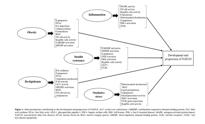
Inflammation, insulin resistance, oxidative stress, obesity and dyslipidemia are the most important factors involved in the pathogenesis of NAFLD (Figure 1). Inflammation, can contribute to the development of NAFLD through increasing the activity of kupffer cells and hepatic stellate cells (HSCs), stimulating nuclear factor κ-B (NF-κB) and mitogen-activated protein kinase (MAPK) pathways, increasing the levels of chemokines as well as toll-like receptors (TLRs), and inducing mitochondrial dysfunction (Buzzetti et al., 2016, Sangouni et al., 2019). The evidence revealed a significant reduction of insulin sensitivity in the muscle, liver and adipose tissue of patients with NAFLD (Lomonaco et al., 2012). Also, insulin resistance can increase the activity of Jun N-terminal kinase (JNK), IκB kinase (IKK), sterol regulatory element-binding proteins (SREBPs), and carbohydrate-responsive element-binding protein (ChREBP), reduce the levels of glucagon-like peptide 1 (GLP-1), and stimulate hepatic de novo lipogenesis (Gaggini et al., 2013, Sangouni and Ghavamzadeh, 2019, Sangouni et al., 2019). By increasing the activity of kupffer cells, HSCs and myeloperoxidase, inducing mitochondrial dysfunction, stimulating TLRs gene expression, and increasing the levels of reactive oxygen species (ROS), oxidative stress can contribute to the pathogenesis of NAFLD (Ashraf and Sheikh, 2015, Sangouni et al., 2019). Moreover, obesity can increase the levels of chemokines, adipocytokines and free fatty acids (FFAs), stimulate the inflammatory pathways, and induce the main pathways of oxidative stress as well as insulin resistance (Polyzos et al., 2019). The hepatic fat accumulation (due to abnormal lipid metabolism or dyslipidemia) is another factor involved in the development and progression of steatosis. Dyslipidemia can reduce the levels of adiponectin, increase the activity of fatty acid synthase (FAS), acetyl-coA carboxylase (ACC), NF-κB and SREBP, increase FFAs and adipokines production, decrease fat oxidation, stimulate hepatic de novo lipogenesis, and induce the pathways of both inflammation and oxidative stress (Ress and Kaser, 2016, Sangouni and Ghavamzadeh, 2019).
Inflammation, insulin resistance, oxidative stress, obesity and dyslipidemia are the most important factors involved in the pathogenesis of NAFLD (Figure 1). Inflammation, can contribute to the development of NAFLD through increasing the activity of kupffer cells and hepatic stellate cells (HSCs), stimulating nuclear factor κ-B (NF-κB) and mitogen-activated protein kinase (MAPK) pathways, increasing the levels of chemokines as well as toll-like receptors (TLRs), and inducing mitochondrial dysfunction (Buzzetti et al., 2016, Sangouni et al., 2019). The evidence revealed a significant reduction of insulin sensitivity in the muscle, liver and adipose tissue of patients with NAFLD (Lomonaco et al., 2012). Also, insulin resistance can increase the activity of Jun N-terminal kinase (JNK), IκB kinase (IKK), sterol regulatory element-binding proteins (SREBPs), and carbohydrate-responsive element-binding protein (ChREBP), reduce the levels of glucagon-like peptide 1 (GLP-1), and stimulate hepatic de novo lipogenesis (Gaggini et al., 2013, Sangouni and Ghavamzadeh, 2019, Sangouni et al., 2019). By increasing the activity of kupffer cells, HSCs and myeloperoxidase, inducing mitochondrial dysfunction, stimulating TLRs gene expression, and increasing the levels of reactive oxygen species (ROS), oxidative stress can contribute to the pathogenesis of NAFLD (Ashraf and Sheikh, 2015, Sangouni et al., 2019). Moreover, obesity can increase the levels of chemokines, adipocytokines and free fatty acids (FFAs), stimulate the inflammatory pathways, and induce the main pathways of oxidative stress as well as insulin resistance (Polyzos et al., 2019). The hepatic fat accumulation (due to abnormal lipid metabolism or dyslipidemia) is another factor involved in the development and progression of steatosis. Dyslipidemia can reduce the levels of adiponectin, increase the activity of fatty acid synthase (FAS), acetyl-coA carboxylase (ACC), NF-κB and SREBP, increase FFAs and adipokines production, decrease fat oxidation, stimulate hepatic de novo lipogenesis, and induce the pathways of both inflammation and oxidative stress (Ress and Kaser, 2016, Sangouni and Ghavamzadeh, 2019).
Gut microbiota
Gut microbiota as a metabolic organ refers to a community of microbes in the gut (Gomaa, 2020). Bacteria, archaea, fungi, phages and viruses are the microorganisms that live in the human gut (Gomaa, 2020). The main bacteria phyla in the human gut are Firmicutes, Bacteroidetes, Proteobacteria, Verrucomicrobia and Actinobacteria (Bull and Plummer, 2014). Firmicutes and Proteobacteria exist mainly in the proximal of GI tract, while Bacteriodetes, Verrucomicrobia are the main phyla in the distal of GI tract (Gu et al., 2013). In addition, Bacteroides, Bifidobacterium, Eubacterium, Clostridium, Peptococcus, Ruminococcus, Peptostreptococcus and Faecalibacterium are the most abundant bacteria genera (Shapira, 2016). Imbalance in the microbial community in terms of qualitative and quantitative changes, distribution, and metabolic activity refers to altered gut microbiota (dysbiosis) (Bull and Plummer, 2014). Factors like age, mode of delivery at birth, exposure to medical treatments, pH of intestinal lumen, diet, physical activity, circadian activity, geographical localization, prebiotics, probiotics and synbiotics can change the gut microbiota composition and distributions (Bull and Plummer, 2014, Clemente et al., 2012). Furthermore, dysbiosis has a critical role in the pathogenesis of atherosclerosis, gastrointestinal diseases, cancers, autoimmune diseases, allergic diseases and T2DM (Bull and Plummer, 2014, Clemente et al., 2012). The recent evidence suggested that gut microbiota plays an important role in the pathogenesis of NAFLD (Leung et al., 2016). In this narrative review, the authors focused on the most recent advances regarding the role of gut microbiota in the pathogenesis of NAFLD. In addition, the mechanisms and clinical results obtained from experimental and clinical studies investigating the synbiotic efficacy in NAFLD were reviewed.
Materials and Methods
PubMed, Scopus, and Web of Science databases were searched in this narrative review to identify the randomized controlled trials (RCTs) which evaluate the effects of synbiotics on hepatic steatosis, fibrosis and liver enzymes in patients with NAFLD. It was sued the following keywords keywords: "Non-alcoholic fatty liver disease'', "Non-alcoholic steatohepatitis'', "Synbiotic'', "Steatosis'', "Fibrosis", "Liver enzyme'', "Aspartate aminotransferase", "Alanine aminotransferase", "Gamma-glutamyltransferase", and "Alkaline phosphatase'' were reviewed. Eight studies investigating the effects of synbiotics on steatosis, fibrosis or liver enzymes in subjects with NAFLD were also included.
In addition, PubMed, Scopus, and Web of Science databases were searched to identify experimental studies, investigating the main mechanisms and pathways that link the gut microbiota to the NAFLD, and the important mechanisms and pathways were completely extracted by careful reading of various related studies. It should be noted that studies published after 2010 were selected.
In addition, PubMed, Scopus, and Web of Science databases were searched to identify experimental studies, investigating the main mechanisms and pathways that link the gut microbiota to the NAFLD, and the important mechanisms and pathways were completely extracted by careful reading of various related studies. It should be noted that studies published after 2010 were selected.
Results
Gut microbiota and NAFLD
The role of dysbiosis in the pathogenesis of NAFLD has been revealed (Leung et al., 2016). About 90% of all gut microbial cells belongs to the phyla Firmicutes and Bacteroidetes (Bull and Plummer, 2014). Patients with NAFLD compared to the healthy controls have higher concentrations of the bacteria belonging to the phylum Firmicutes (Raman et al., 2013). The abundance of Porphyromonas, Campylobacter, Staphylococcus, Parabacteroides, Dialister and Ruminococcus is higher in subjects with obesity compared to the healthy people (Le Chatelier et al., 2013). In addition, lower concentrations of Lactobacillus, Bifidobacterium, Faecalibacterium, Akkermansia, Methanobrevibacter and Coprococcus in subjects with obesity were found (Gomaa, 2020, Le Chatelier et al., 2013). Patients with NAFLD have increased concentrations of Dorea and Ruminococcus (Del Chierico et al., 2017). An inverse relationship between concentration of Bifidobacterium and levels of alanine aminotransferase levels was found in subjects with higher risk of NAFLD (Karlsson et al., 2012). Moreover, a recent investigation showed that the concentration of Firmicutes was more in mild or moderate NAFLD; but, the patients with advanced fibrosis have higher concentration of Proteobacteria (Loomba et al., 2017). The abundance of bacteria in the gut in patients with NAFLD and healthy people are summarized in Table 1. Dysbiosis can induce hepatic steatosis and NASH through following pathways and mechanisms (Table 2).
| Table 1. The bacteria abundance in the gut; Patients with NAFLD compared to the healthy people. | |
| Bacteria with increased abundance | Bacteria with decreased abundance |
| Robinsoniella Porphyromonas Campylobacter Staphylococcus Parabacteroides Dialister Ruminococcus Dorea Clostridium Klebsiella pneumoniae |
Lactobacillus Bifidobacterium Faecalibacterium Akkermansia Methanobrevibacter Coprococcus Eubacterium |
| NAFLD: non-alcoholic fatty liver disease | |
| Table 2. Main pathways and mechanisms that link dysbiosis to the NAFLD. | |
| Pathway | Mechanisms |
| Gut permeability | ↑LPS, ↑SIBO, disrupting epithelial tight junctions, ↑degradation of the mucus layer, ↓production of mucus, changing the intestinal barrier, ↑leptin, ↓adiponectin, ↑SREBPs activation, ↑endotoxemia, ↑plasminogen activator inhibitor 1, ↑hepatic TLR4 expression, ↓GLP2, ↑insulin resistance, ↑inflammatory pathways. |
| Stimulation of obesity | ↑GPR43 activation, ↑small intestinal villi density, ↑ChREBPs activation, ↑hepatic lipogenesis, ↓lipolysis, ↓FIAF expression, ↓GLP1, ↑LPL activity. |
| Stimulation of inflammation | ↑LPS, ↑TLR4, ↑NF-κβ pathways activation, ↑TNF-α, ↑HSCs activation, ↑IL-6, ↑MAPK pathways activation, ↑kupffer cells activity, ↑inflammasomes, ↓GLP1, ↑JNK pathways activation, ↓claudin expression, ↓occludin expression, ↓ZO-1 expression |
| Insulin resistance | ↑TLR4, ↑LPS, changing immunity response, ↑endotoxemia, ↑IRS 1 serine phosphorylation, ↓GLP1, ↑IKK, ↑JNK pathways activation, ↓occludin expression, ↓ZO-1 expression. |
| Dietary choline metabolism | ↑dimethylamine, ↑trimethylamine, ↑TMAO, ↑NF-κβ pathways activation, ↑TNF-α, ↑IL-6, ↑MAPK pathways activation, ↑kupffer cells activity, ↑HSCs activation. |
| Bile acid metabolism | ↓FXR activation, GPBAR1 dysregulation, ↓GLP1, ↑hepatic de novo lipogenesis, ↑VLDL, ↑NF-κβ pathways activation, ↑ROS production, ↑lipid peroxidation, ↑kupffer cells activity. |
| Ethanol production | ↑kupffer cells activity, ↑TNF-α, ↑IL-6, ↑IL-1β, ↑ROS production in the liver, ↑hepatic TLR4, ↑liver enzymes levels, ↑passage of gut-derived endotoxins into portal system, ↑gut permeability. |
| ChREBP: Carbohydrate-responsive element-binding protein; FIAF: Fasting-induced adipocyte factor; FXR: Farnesoid X receptor; GLP: glucagon-like peptide; GPBAR1: G-protein coupled bile acid receptor 1; GPR43: G protein-coupled receptor 43; HSCs: Hepatic stellate cells; IKK: IκB kinase; IL-6: Interleukin-6; IL-1β: Interleukin-1β; IRS1: Insulin receptor substrate 1; JNK: Jun N-terminal kinase; LPL: lipoprotein lipase; LPS: lipopolysaccharide; MAPK: mitogen-activated protein kinase; NAFLD: non-nlcoholic fatty liver disease; NF-κB: Nuclear factor κ-B; ROS: Reactive oxygen species; SIBO: Small intestinal bacterial overgrowth; SREBPs: Sterol regulatory element-bindings; TG: Triglyceride; TLR4: Toll-like receptor 4; TMAO: Trimethylamine N-Oxide; TNF-α: Tumor necrosis factor-α; VLDL: Very low-density lipoprotein; ZO-1: Zonula occludens-1. |
|
Gut permeability
Gut permeability is an important mechanism involved in the pathogenesis of NAFLD. It refers to the decreased integrity of junctions in the gut epithelial wall that leads to an increase in material translocation from the lumen into the bloodstream, other organs, and adipose tissue (Kolodziejczyk et al., 2019). A direct correlation between NAFLD with gut permeability related to the small intestinal bacterial overgrowth (SIBO) and disrupted tight junctions was also observed (Fukunishi et al., 2014, Miele et al., 2009). Lipopolysaccharides (LPS), which are found in the outer membrane of gram-negative bacteria, can enhance SIBO and disrupt tight junctions (Fukunishi et al., 2014, Miele et al., 2009). Moreover, LPS decreases plasma adiponectin, enhances plasma leptin, and triggers expression of liver SREBP-1c (Fukunishi et al., 2014). Dysbiosis decreases the GLP-2 secretion, disrupts tight junctions, and subsequently, increases the gut permeability(Tseng and Wu, 2019). Disrupted tight junctions can enhance the endotoxemia (Miele et al., 2009, Volynets et al., 2012). Moreover, endotoxemia enhances the expression of the plasma plasminogen activator inhibitor 1 and hepatic TLR4, which can stimulate the development of inflammation and insulin resistance (Alisi et al., 2010, Volynets et al., 2012).
Stimulation of obesity
It has been revealed that the abundance of Bifidobacterium, Oscillospira, Erwinia, Succinivibrio and Alistipes is higher in subjects with normal weight compared to obese individuals (Ignacio et al., 2016, Tseng and Wu, 2019). In addition, a significant relationship between bacteria species such as Blautia hydrogenotorophica, Coprococcus catus, Ruminococcus bromii and Ruminococcus obeum and obesity was found (Kasai et al., 2015). The close link between gut microbiota and obesity has also been confirmed. The gut microbiota as an important regulator of the energy harvest can enhance the density of small intestinal villi, produce short-chain fatty acids (SCFAs), induce gut motility, and increase fat deposition (Delzenne and Cani, 2011, Gomaa, 2020). SCFAs reduce the glucagon-like peptide 1 (GLP1) secretion and activate the G-protein coupled receptors 43 (GPR43), inhibit lipolysis and increase fat storage (Morrison and Preston, 2016). SCFAs can activate the ChREBP and increase the expression of proteins which enhance hepatic lipogenesis (Morrison and Preston, 2016). On the other hand, fasting-induced adipocyte factor (FIAF) stimulates changes in the expression of genes controlling energy homeostasis, reduces the lipoprotein lipase (LPL) activity in the adipose tissue, enhances fatty acid oxidation, upregulates uncoupling proteins, and can potentially reduce the amount of fat storage (Arslan, 2014, Shen et al., 2013). Moreover, dysbiosis decreases the synthesis and secretion of FIAF, triggers activity of LPL and increases fat storage in the liver (Arslan, 2014, Delzenne and Cani, 2011, Shen et al., 2013).
Stimulation of inflammation
Dysbiosis induces low-grade inflammation, an important factor in the development of obesity and NAFLD. The evidence suggested that TLR4 contributes to the development of NAFLD (Aron-Wisnewsky et al., 2013, Canfora et al., 2019). The kupffer cells, which can trigger liver expression of TLR4, play a critical role in the liver inflammation by producing inflammatory cytokines, chemokines, and ROS (Aron-Wisnewsky et al., 2013, Paik et al., 2003). LPS increases kupffer cells activity and tumor necrosis factor-α (TNF-α). Also, HSCs trigger endotoxemia and inflammatory pathways in the liver (Aron-Wisnewsky et al., 2013, Paik et al., 2003). The endotoxemia resulted from dysbiosis induces insulin resistance and inflammation via pathways related to the LPS-TLR4, and stimulates JNK and NF-κB pathways (Henao-Mejia et al., 2012, Verdam et al., 2011). Eventually, these actions increase inflammasomes and pro-inflammatory cytokines (Henao-Mejia et al., 2012).
Stimulation of insulin resistance
Dysbiosis increases the ghrelin secretion, and subsequently increases the energy storage and insulin resistance (Caricilli and Saad, 2013, Perry et al., 2016). TLR4 is involved in the development of insulin resistance by activating LPS, increasing gene expression of liver SREBP-1c and decreasing level of adiponectin (Caricilli and Saad, 2013, Perry et al., 2016).
Dysbiosis inhibits the expression of tight junction proteins occludin and zonula occludens-1 (ZO-1), increases intestinal permeability, and enhances endotoxemia (Lee et al., 2020). Endotoxemia derived from the gut can trigger mechanisms and pathways related to the LPS-TLR4, and activate the JNK and NF-kB pathways (Lee et al., 2020, Verdam et al., 2011). Additionally, TLR4 activates IKK as well as JNK and reduces insulin receptor substrate 1 (IRS-1) serine phosphorylation and insulin sensitivity (Kolodziejczyk et al., 2019, Lee et al., 2020).
Dysbiosis inhibits the expression of tight junction proteins occludin and zonula occludens-1 (ZO-1), increases intestinal permeability, and enhances endotoxemia (Lee et al., 2020). Endotoxemia derived from the gut can trigger mechanisms and pathways related to the LPS-TLR4, and activate the JNK and NF-kB pathways (Lee et al., 2020, Verdam et al., 2011). Additionally, TLR4 activates IKK as well as JNK and reduces insulin receptor substrate 1 (IRS-1) serine phosphorylation and insulin sensitivity (Kolodziejczyk et al., 2019, Lee et al., 2020).
Dietary choline metabolism
Choline has an important role in the hepatic biogenesis of phosphatidylcholine and regulation of S-adenosyl methionine cycle(Kolodziejczyk et al., 2019); through increasing hepatic biogenesis of phosphatidylcholine, Choline is involved in very low-density lipoprotein (VLDL) production, and hepatic lipid export (Aron-Wisnewsky et al., 2013). Meat products, dairy products, soybeans, fishes, nuts and whole grains are dietary sources of choline, while gut microbiome is an important endogenous source of choline (Russell et al., 2013). Choline deficiency is associated with NAFLD, and dysbiosis reduces choline bioavailability by producing toxic metabolites such as dimethylamine and trimethylamine from dietary choline (Russell et al., 2013, Wang et al., 2011). Species such as Anaerococcus hydrogenalis, Clostridium asparagiforme, Clostridium hathewayi, Clostridium sporogenes, Escherichia fergusonii, Proteus penneri, Providencia rettgeri and Edwardsiella tarda are involved in the conversion of choline to trimethylamine N-oxide (TMAO) (Romano et al., 2015). TMAO derived from dimethylamine and trimethylamine in the liver triggers inflammatory pathways (Wang et al., 2011). Furthermore, choline deficiency stimulates NF-κβ pathways and increases the hepatic levels of inflammatory markers such as TNF-α and IL-6, activating MAPK pathways as well as the activity of kupffer cells and HSCs (Wang et al., 2011). High levels of TMAO increase the abundance of Clostridium (Spencer et al., 2011). Moreover, mutation in phosphatidylethanolamine N-methyltransferase (PEMT) gene, which is involved in the formation of endogenous choline, can lead to the progression of NAFLD (Kolodziejczyk et al., 2019, Leung et al., 2016).
Bile acid metabolism
The secondary bile acids are made by the gut microbiota from cholesterol-derived primary bile acids produced in the liver (Ferslew et al., 2015). Farnesoid X receptor (FXR) is the receptor of bile acids (Brighton et al., 2015, Trauner et al., 2010). Accordingly, bile acids activate FXR and G-protein coupled bile acid receptor 1 (GPBAR1), and subsequently activate FXR and GPBAR1 pathways, which are the important regulators of the hepatic de novo lipogenesis (Brighton et al., 2015, Hylemon et al., 2009). The level of hydrophobic and cytotoxic BAs species is higher in patients with NAFLD (Ferslew et al., 2015). The enzymes involved in the conversion of deconjugated BAs to secondary BAs are also expressed throughout the gut microbiota (Ridlon et al., 2013). Dysbiosis disrupts enterohepatic circulation and alters structure of bile acids (Swann et al., 2011); it changes expression and function of FXR, secretion of bile acids and contributes to the hepatic steatosis, inflammation and oxidative stress (Kolodziejczyk et al., 2019, Swann et al., 2011).
Ethanol production
Gut microbiota produces ethanol, and the ethanol is transported to the liver by portal system (Leung et al., 2016). Ethanol and its main hepatotoxic metabolites including acetaldehyde and acetate trigger kupffer cells to produce inflammatory markers like TNF-α (Arslan, 2014, Gustot et al., 2006). Ethanol induces the production of ROS by hepatocytes, increases the levels of liver enzymes, and induces the expression of TLRs (Arslan, 2014, Gustot et al., 2006). The abundance of ethanol-producer bacteria such as Proteobacteria, Enterobacteriaceae, and Escherichia is higher in subjects with NASH (Zhu et al., 2013). In addition, high-ethanol-Producing Klebsiella pneumoniae is abundant in patients with NAFLD. Klebsiella pneumoniae is strongly associated with NAFLD, and transplant of Klebsiella pneumoniae causes NAFLD (Forsyth et al., 2011, Zhu et al., 2013). Finally, ethanol enhances the gut permeability and increases transfer of gut-derived endotoxins into the liver (Forsyth et al., 2011).
Synbiotics
Synbiotic is a synergistic combination of prebiotic and probiotic (Vyas and Ranganathan, 2012). Prebiotics as the indigestible carbohydrates have a beneficial effect on host health by modifying gut microbiota composition, distribution and activity (Vyas and Ranganathan, 2012). Probiotics are the nonpathogenic living microorganisms, and in sufficient quantities, they can improve the gut microbiota dysbiosis (Vyas and Ranganathan, 2012). Lacbobacilli, Bifidobacteria spp, S. boulardii, and B. coagulans are the most common probiotic strains in the synbiotics formulations (Pandey et al., 2015). Fructooligosaccharide (FOS), yloseoligosaccharide (XOS), inulin, and prebiotics from natural sources like chicory are the main prebiotics that are used in the formulations of synbiotics (Pandey et al., 2015). Synbiotics can stimulate the growth of health-promoting bacteria, activate the metabolism of one health-promoting, improve the survival of probiotics and live microbial implantation in the GI tract (Cencic and Chingwaru, 2010, Pandey et al., 2015). Presumably, synbiotics were developed to attenuate the difficulties related to probiotics survival. The beneficial effect of synbiotics on survival of probiotic bacteria during passage through the upper gastrointestinal tract has been observed as well (Cencic and Chingwaru, 2010, Pandey et al., 2015). Synbiotics by improving the implantation of beneficial bacteria in the gut as well as stimulating the growth of probiotics are involved in maintaining intestinal homeostasis and health (Cencic and Chingwaru, 2010, Pandey et al., 2015, Vyas and Ranganathan, 2012).
Synbiotics and NAFLD
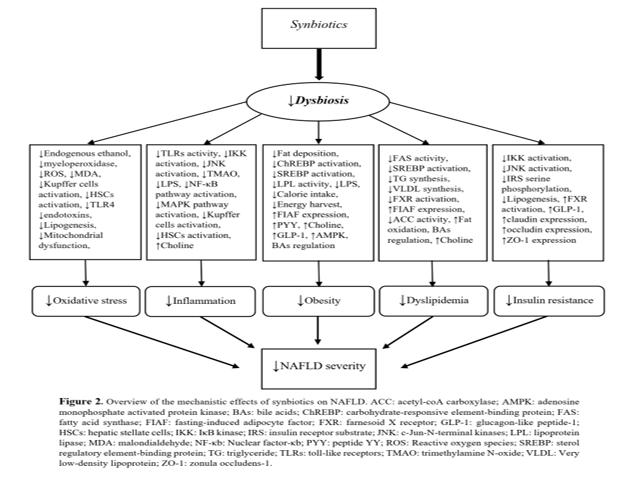
Pathways and mechanisms: A NAFLD model revealed that synbiotics can regulate gut microbiota, and reduce the severity of steatosis as well as fibrosis (Cortez-Pinto et al., 2016). The evidence showed that synbiotics decrease endogenous ethanol, levels of TMAO and myeloperoxidase secretion, attenuate the production of ROS as well as lipid peroxidation products, decrease the activity of kupffer cells, HSCs, IKK and JNK, downregulate production of liver inflammatory markers, reduce the activity of NF-κB and MAPK pathways, decrease the mitochondrial dysfunction, increase levels of choline, decrease the expression of TLRs, and subsequently, improve the main causes of NAFLD such as inflammation and oxidative stress (Raso et al., 2014, Sangouni and Ghavamzadeh, 2019). In addition, synbiotics can regulate bile acids; attenuate lipogenesis and fat deposition; decrease levels of LPS and endotoxin, ChREBP and SREBP activity, and TG and VLDL synthesis; , , increase fat oxidation, peptide YY (PYY), GLP-1 and adenosine monophosphate activated protein kinase (AMPK), FIAF expression, and claudin, and occludin and ZO-1 expression; reduce FAS, ACC, IKK, JNK and FXR activity, LPL, calorie intake and energy harvest, and IRS serine phosphorylation, and ultimately, improve gut permeability. They all lead to an improvement in obesity, insulin resistance, and dyslipidemia improvement (Sangouni and Ghavamzadeh, 2019). A narrative review confirmed the beneficial effects of synbiotics on main risk factors of NAFLD such as insulin resistance, obesity, inflammation and oxidative stress (Sangouni and Ghavamzadeh, 2019). The pathways and mechanisms related to the synbiotics efficacy in NAFLD are represented in Figure 2.
Synbiotic and hepatic steatosis and fibrosis
Several RCTs have examined the effect of synbiotics supplementation on hepatic steatosis and fibrosis in patients with NAFLD (Table 3). Mofidi et al. (Mofidi et al., 2017) reported a significant reduction in steatosis and fibrosis after 28 weeks of synbiotics supplementation. Moreover, the study of Malaguarnera et al. (Malaguarnera et al., 2012) showed an improvement in steatosis and NASH activity score after synbiotics administration; however, synbiotics supplementation could not change fibrosis. In addition, the studies of Asgharian et al. (Asgharian et al., 2016), Bakhshimoghaddam et al. (Bakhshimoghaddam et al., 2018), and Ferolla et al. (Ferolla et al., 2016) demonstrated a significant reduction in the severity of steatosis in synbiotic group. Eslamparast et al. (Eslamparast et al., 2014) also showed a significant reduction in fibrosis score after synbiotics supplementation for 28 weeks. However, Malaguarnera et al. (Malaguarnera et al., 2012), and Ferolla et al. (Ferolla et al., 2016) found that there is no significant difference between synbiotic and control groups in level of fibrosis after synbiotics supplementation. It seems that synbiotics have a therapeutic effect on hepatic steatosis. However, the clinical results of synbiotics on fibrosis are inconsistent. This inconsistency can be due to the difference of studies in intervention duration. Studies with higher intervention duration showed beneficial effect of synbiotics on hepatic fibrosis.
Synbiotic and liver enzymes
The effect of synbiotics supplementation on liver enzymes in patients with NAFLD was investigated in several RCTs (Table 3). Eslamparast et al. (Eslamparast et al., 2014) found a significant reduction in levels of liver enzymes including aspartate aminotransferase (AST), alanine aminotransferase (ALT) and gamma-glutamyltransferase (GGT) after 28 weeks; but, alkaline phosphatase (ALP) remained unchanged. The study of Bakhshimoghaddam et al. showed a greater decrease in AST, ALT, GGT and ALP serum levels in synbiotic group compared to the control group after 28 weeks. In addition, in the study by Ekhlasi et al. (Ekhlasi et al., 2017), a significant reduction in levels of ALT, AST and ALP was found in the synbiotic group. Moreover, the studies of Malaguarnera et al. (Malaguarnera et al., 2012), and Sayari et al. (Sayari et al., 2018) found a significant decrease in AST in synbiotic group after intervention period; however, the levels of ALT did not change. Mofidi et al.’s study was conducted among lean patients with NAFLD and a significant difference was found between synbiotic and placebo in AST levels; but, there was no significant change in the levels of ALT and GGT after supplementation for 28 weeks. The studies of Asgharian et al. (Asgharian et al., 2016) and Ferolla et al. (Ferolla et al., 2016) reported that the levels of liver enzymes remained unchanged after synbiotic supplementation. In general, the studies with longer intervention durations showed a greater improvement of liver enzymes.
Conclusions
Modifying gut microbiota composition and distribution can be considered as a therapeutic strategy in NAFLD. The studies investigating the effects of synbiotics on liver enzymes and hepatic steatosis showed promising results. However, the clinical results related to synbiotics and fibrosis are inconsistent. To clarify the clinical effects of synbiotics on fibrosis, more investigations are needed.
Authors' contributions
All the authors have contributed to the various stages of this work.
Conflict of interests
The authors declared no competing interests.
References
Alisi A, Manco M, Devito R, Piemonte F & Nobili V 2010. Endotoxin and plasminogen activator inhibitor-1 serum levels associated with nonalcoholic steatohepatitis in children. Journal of pediatric gastroenterology and nutrition. 50 (6): 645-649.
Aron-Wisnewsky J, Gaborit B, Dutour A & Clement K 2013. Gut microbiota and non-alcoholic fatty liver disease: new insights. Clinical microbiology and infection. 19 (4): 338-348.
Arslan N 2014. Obesity, fatty liver disease and intestinal microbiota. World journal of gastroenterology. 20 (44): 16452.
Asgharian A, Askari G, Esmailzade A, Feizi A & Mohammadi V 2016. The effect of symbiotic supplementation on liver enzymes, C-reactive protein and ultrasound findings in patients with non-alcoholic fatty liver disease: a clinical trial. International journal of preventive medicine. 7 (1): 59.
Ashraf N & Sheikh T 2015. Endoplasmic reticulum stress and oxidative stress in the pathogenesis of non-alcoholic fatty liver disease. Free radical research. 49 (12): 1405-1418.
Bakhshimoghaddam F, Shateri K, Sina M, Hashemian M & Alizadeh M 2018. Daily consumption of synbiotic yogurt decreases liver steatosis in patients with nonalcoholic fatty liver disease: a randomized controlled clinical trial. Journal of nutrition. 148 (8): 1276-1284.
Brighton CA, et al. 2015. Bile acids trigger GLP-1 release predominantly by accessing basolaterally located G protein–coupled bile acid receptors. Endocrinology. 156 (11): 3961-3970.
Bull M & Plummer N 2014. Part 1: The human gut microbiome in health and disease. Integrated medicine. 13 (6): 17-22.
Buzzetti E, Pinzani M & Tsochatzis EA 2016. The multiple-hit pathogenesis of non-alcoholic fatty liver disease (NAFLD). Metabolism. 65 (8): 1038-1048.
Canfora EE, Meex RC, Venema K & Blaak EE 2019. Gut microbial metabolites in obesity, NAFLD and T2DM. Nature reviews endocrinology. 15 (5): 261-273.
Caricilli AM & Saad MJ 2013. The role of gut microbiota on insulin resistance. Nutrients. 5 (3): 829-851.
Cencic A & Chingwaru W 2010. The role of functional foods, nutraceuticals, and food supplements in intestinal health. Nutrients. 2 (6): 611-625.
Chalasani N, et al. 2012. The diagnosis and management of non‐alcoholic fatty liver disease: Practice guideline by the American Association for the study of liver diseases, American College of Gastroenterology, and the American Gastroenterological Association. Hepatology. 55 (6): 2005-2023.
Clemente JC, Ursell LK, Parfrey LW & Knight R 2012. The impact of the gut microbiota on human health: an integrative view. Cell. 148 (6): 1258-1270.
Cortez-Pinto H, et al. 2016. Microbiota modulation with synbiotic decreases liver fibrosis in a high fat choline deficient diet mice model of non-alcoholic steatohepatitis (NASH). GE Portuguese journal of gastroenterology. 23 (3): 132-141.
Del Chierico F, et al. 2017. Gut microbiota profiling of pediatric nonalcoholic fatty liver disease and obese patients unveiled by an integrated meta‐omics‐based approach. Hepatology. 65 (2): 451-464.
Delzenne NM & Cani PD 2011. Interaction between obesity and the gut microbiota: relevance in nutrition. Annual review of nutrition. 31 (1): 15-31.
Ekhlasi G, et al. 2017. Effects of symbiotic and vitamin E supplementation on blood pressure, nitric oxide and inflammatory factors in non-alcoholic fatty liver disease. EXCLI journal. 16: 278.
Elizabeth LY, Schwimmer JB & Lavine JE 2010. Non-alcoholic fatty liver disease: epidemiology, pathophysiology, diagnosis and treatment. Paediatrics and child health. 20 (1): 26-29.
Eslamparast T, et al. 2014. Synbiotic supplementation in nonalcoholic fatty liver disease: a randomized, double-blind, placebo-controlled pilot study. American journal of clinical nutrition. 99 (3): 535-542.
Ferolla SM, et al. 2016. Beneficial effect of synbiotic supplementation on hepatic steatosis and anthropometric parameters, but not on gut permeability in a population with nonalcoholic steatohepatitis. Nutrients. 8 (7): 397.
Ferslew BC, et al. 2015. Altered bile acid metabolome in patients with nonalcoholic steatohepatitis. Digestive diseases and sciences. 60: 3318-3328.
Forsyth CB, Tang Y, Shaikh M, Zhang L & Keshavarzian A 2011. Role of snail activation in alcohol‐induced iNOS‐mediated disruption of intestinal epithelial cell permeability. Alcoholism: Clinical and experimental research. 35 (9): 1635-1643.
Fukunishi S, et al. 2014. Lipopolysaccharides accelerate hepatic steatosis in the development of nonalcoholic fatty liver disease in Zucker rats. Journal of clinical biochemistry and nutrition. 54 (1): 39-44.
Gaggini M, et al. 2013. Non-alcoholic fatty liver disease (NAFLD) and its connection with insulin resistance, dyslipidemia, atherosclerosis and coronary heart disease. Nutrients. 5 (5): 1544-1560.
Gomaa EZ 2020. Human gut microbiota/microbiome in health and diseases: a review. Antonie Van Leeuwenhoek. 113 (12): 2019-2040.
Gu S, et al. 2013. Bacterial community mapping of the mouse gastrointestinal tract. PloS one. 8 (10): e74957.
Gustot T, et al. 2006. Differential liver sensitization to toll‐like receptor pathways in mice with alcoholic fatty liver. Hepatology. 43 (5): 989-1000.
Henao-Mejia J, et al. 2012. Inflammasome-mediated dysbiosis regulates progression of NAFLD and obesity. Nature. 482 (7384): 179-185.
Hylemon PB, et al. 2009. Bile acids as regulatory molecules. Journal of lipid research. 50 (8): 1509-1520.
Ignacio A, et al. 2016. Correlation between body mass index and faecal microbiota from children. Clinical microbiology and infection. 22 (3): 258. e251-258. e258.
Karlsson CL, et al. 2012. The microbiota of the gut in preschool children with normal and excessive body weight. Obesity. 20 (11): 2257-2261.
Kasai C, et al. 2015. Comparison of the gut microbiota composition between obese and non-obese individuals in a Japanese population, as analyzed by terminal restriction fragment length polymorphism and next-generation sequencing. BMC gastroenterology. 15: 1-10.
Kolodziejczyk AA, Zheng D, Shibolet O & Elinav E 2019. The role of the microbiome in NAFLD and NASH. EMBO molecular medicine. 11 (2): e9302.
Le Chatelier E, et al. 2013. Richness of human gut microbiome correlates with metabolic markers. Nature. 500 (7464): 541-546.
Lee CJ, Sears CL & Maruthur N 2020. Gut microbiome and its role in obesity and insulin resistance. Annals of the New York Academy of Sciences. 1461 (1): 37-52.
Leung C, Rivera L, Furness JB & Angus PW 2016. The role of the gut microbiota in NAFLD. Nature reviews gastroenterology & hepatology. 13 (7): 412-425.
Lomonaco R, et al. 2012. Effect of adipose tissue insulin resistance on metabolic parameters and liver histology in obese patients with nonalcoholic fatty liver disease. Hepatology. 55 (5): 1389-1397.
Loomba R, et al. 2017. Gut microbiome-based metagenomic signature for non-invasive detection of advanced fibrosis in human nonalcoholic fatty liver disease. Cell metabolism. 25 (5): 1054-1062. e1055.
Malaguarnera M, et al. 2012. Bifidobacterium longum with fructo-oligosaccharides in patients with non alcoholic steatohepatitis. Digestive diseases and sciences. 57: 545-553.
Miele L, et al. 2009. Increased intestinal permeability and tight junction alterations in nonalcoholic fatty liver disease. Hepatology. 49 (6): 1877-1887.
Mofidi F, et al. 2017. Synbiotic supplementation in lean patients with non-alcoholic fatty liver disease: a pilot, randomised, double-blind, placebo-controlled, clinical trial. British Journal of Nutrition. 117 (5): 662-668.
Morrison DJ & Preston T 2016. Formation of short chain fatty acids by the gut microbiota and their impact on human metabolism. Gut microbes. 7 (3): 189-200.
Paik YH, et al. 2003. Toll‐like receptor 4 mediates inflammatory signaling by bacterial lipopolysaccharide in human hepatic stellate cells. Hepatology. 37 (5): 1043-1055.
Pandey KR, Naik SR & Vakil BV 2015. Probiotics, prebiotics and synbiotics-a review. Journal of food science and technology. 52: 7577-7587.
Perry RJ, et al. 2016. Acetate mediates a microbiome–brain–β-cell axis to promote metabolic syndrome. Nature. 534 (7606): 213-217.
Polyzos SA, Kountouras J & Mantzoros CS 2019. Obesity and nonalcoholic fatty liver disease: From pathophysiology to therapeutics. Metabolism. 92: 82-97.
Raman M, et al. 2013. Fecal microbiome and volatile organic compound metabolome in obese humans with nonalcoholic fatty liver disease. Clinical gastroenterology and hepatology. 11 (7): 868-875. e863.
Raso GM, et al. 2014. Effects of a Lactobacillus paracasei B21060 based synbiotic on steatosis, insulin signaling and toll-like receptor expression in rats fed a high-fat diet. Journal of nutritional biochemistry. 25 (1): 81-90.
Ress C & Kaser S 2016. Mechanisms of intrahepatic triglyceride accumulation. World journal of gastroenterology. 22 (4): 1664.
Ridlon JM, Alves JM, Hylemon PB & Bajaj JS 2013. Cirrhosis, bile acids and gut microbiota: unraveling a complex relationship. Gut microbes. 4 (5): 382-387.
Romano KA, Vivas EI, Amador-Noguez D & Rey FE 2015. Intestinal microbiota composition modulates choline bioavailability from diet and accumulation of the proatherogenic metabolite trimethylamine-N-oxide. MBio. 6 (2): 10.1128/mbio. 02481-02414.
Russell WR, Hoyles L, Flint HJ & Dumas M-E 2013. Colonic bacterial metabolites and human health. Current opinion in microbiology. 16 (3): 246-254.
Sangouni AA & Ghavamzadeh S 2019. A review of synbiotic efficacy in non-alcoholic fatty liver disease as a therapeutic approach. Diabetes & metabolic syndrome: Clinical research & reviews. 13 (5): 2917-2922.
Sangouni AA, Ghavamzadeh S & Jamalzehi A 2019. A narrative review on effects of vitamin D on main risk factors and severity of non-alcoholic fatty liver disease. Diabetes & metabolic syndrome: Clinical research & reviews. 13 (3): 2260-2265.
Sayari S, Neishaboori H & Jameshorani M 2018. Combined effects of synbiotic and sitagliptin versus sitagliptin alone in patients with nonalcoholic fatty liver disease. Clinical and molecular hepatology. 24 (3): 331.
Shapira M 2016. Gut microbiotas and host evolution: scaling up symbiosis. Trends in ecology & evolution. 31 (7): 539-549.
Shen J, Obin MS & Zhao L 2013. The gut microbiota, obesity and insulin resistance. Molecular aspects of medicine. 34 (1): 39-58.
Spencer MD, et al. 2011. Association between composition of the human gastrointestinal microbiome and development of fatty liver with choline deficiency. Gastroenterology. 140 (3): 976-986.
Swann JR, et al. 2011. Systemic gut microbial modulation of bile acid metabolism in host tissue compartments. Proceedings of the national academy of sciences. 108 (supplement_1): 4523-4530.
Trauner M, Claudel T, Fickert P, Moustafa T & Wagner M 2010. Bile acids as regulators of hepatic lipid and glucose metabolism. Digestive diseases. 28 (1): 220-224.
Tseng C-H & Wu C-Y 2019. The gut microbiome in obesity. Journal of the Formosan Medical Association. 118: S3-S9.
Verdam FJ, Rensen SS, Driessen A, Greve JW & Buurman WA 2011. Novel evidence for chronic exposure to endotoxin in human nonalcoholic steatohepatitis. Journal of clinical gastroenterology. 45 (2): 149-152.
Volynets V, et al. 2012. Nutrition, intestinal permeability, and blood ethanol levels are altered in patients with nonalcoholic fatty liver disease (NAFLD). Digestive diseases and sciences. 57: 1932-1941.
Vyas U & Ranganathan N 2012. Probiotics, prebiotics, and synbiotics: gut and beyond. Gastroenterology research and practice. 2012 (1): 872716.
Wang Z, et al. 2011. Gut flora metabolism of phosphatidylcholine promotes cardiovascular disease. Nature. 472 (7341): 57-63.
Wong RJ, et al. 2015. Nonalcoholic steatohepatitis is the second leading etiology of liver disease among adults awaiting liver transplantation in the United States. Gastroenterology. 148 (3): 547-555.
Younossi Z, et al. 2018. Global burden of NAFLD and NASH: trends, predictions, risk factors and prevention. Nature reviews gastroenterology & hepatology. 15 (1): 11-20.
Zhu L, et al. 2013. Characterization of gut microbiomes in nonalcoholic steatohepatitis (NASH) patients: a connection between endogenous alcohol and NASH. Hepatology. 57 (2): 601-609.
Aron-Wisnewsky J, Gaborit B, Dutour A & Clement K 2013. Gut microbiota and non-alcoholic fatty liver disease: new insights. Clinical microbiology and infection. 19 (4): 338-348.
Arslan N 2014. Obesity, fatty liver disease and intestinal microbiota. World journal of gastroenterology. 20 (44): 16452.
Asgharian A, Askari G, Esmailzade A, Feizi A & Mohammadi V 2016. The effect of symbiotic supplementation on liver enzymes, C-reactive protein and ultrasound findings in patients with non-alcoholic fatty liver disease: a clinical trial. International journal of preventive medicine. 7 (1): 59.
Ashraf N & Sheikh T 2015. Endoplasmic reticulum stress and oxidative stress in the pathogenesis of non-alcoholic fatty liver disease. Free radical research. 49 (12): 1405-1418.
Bakhshimoghaddam F, Shateri K, Sina M, Hashemian M & Alizadeh M 2018. Daily consumption of synbiotic yogurt decreases liver steatosis in patients with nonalcoholic fatty liver disease: a randomized controlled clinical trial. Journal of nutrition. 148 (8): 1276-1284.
Brighton CA, et al. 2015. Bile acids trigger GLP-1 release predominantly by accessing basolaterally located G protein–coupled bile acid receptors. Endocrinology. 156 (11): 3961-3970.
Bull M & Plummer N 2014. Part 1: The human gut microbiome in health and disease. Integrated medicine. 13 (6): 17-22.
Buzzetti E, Pinzani M & Tsochatzis EA 2016. The multiple-hit pathogenesis of non-alcoholic fatty liver disease (NAFLD). Metabolism. 65 (8): 1038-1048.
Canfora EE, Meex RC, Venema K & Blaak EE 2019. Gut microbial metabolites in obesity, NAFLD and T2DM. Nature reviews endocrinology. 15 (5): 261-273.
Caricilli AM & Saad MJ 2013. The role of gut microbiota on insulin resistance. Nutrients. 5 (3): 829-851.
Cencic A & Chingwaru W 2010. The role of functional foods, nutraceuticals, and food supplements in intestinal health. Nutrients. 2 (6): 611-625.
Chalasani N, et al. 2012. The diagnosis and management of non‐alcoholic fatty liver disease: Practice guideline by the American Association for the study of liver diseases, American College of Gastroenterology, and the American Gastroenterological Association. Hepatology. 55 (6): 2005-2023.
Clemente JC, Ursell LK, Parfrey LW & Knight R 2012. The impact of the gut microbiota on human health: an integrative view. Cell. 148 (6): 1258-1270.
Cortez-Pinto H, et al. 2016. Microbiota modulation with synbiotic decreases liver fibrosis in a high fat choline deficient diet mice model of non-alcoholic steatohepatitis (NASH). GE Portuguese journal of gastroenterology. 23 (3): 132-141.
Del Chierico F, et al. 2017. Gut microbiota profiling of pediatric nonalcoholic fatty liver disease and obese patients unveiled by an integrated meta‐omics‐based approach. Hepatology. 65 (2): 451-464.
Delzenne NM & Cani PD 2011. Interaction between obesity and the gut microbiota: relevance in nutrition. Annual review of nutrition. 31 (1): 15-31.
Ekhlasi G, et al. 2017. Effects of symbiotic and vitamin E supplementation on blood pressure, nitric oxide and inflammatory factors in non-alcoholic fatty liver disease. EXCLI journal. 16: 278.
Elizabeth LY, Schwimmer JB & Lavine JE 2010. Non-alcoholic fatty liver disease: epidemiology, pathophysiology, diagnosis and treatment. Paediatrics and child health. 20 (1): 26-29.
Eslamparast T, et al. 2014. Synbiotic supplementation in nonalcoholic fatty liver disease: a randomized, double-blind, placebo-controlled pilot study. American journal of clinical nutrition. 99 (3): 535-542.
Ferolla SM, et al. 2016. Beneficial effect of synbiotic supplementation on hepatic steatosis and anthropometric parameters, but not on gut permeability in a population with nonalcoholic steatohepatitis. Nutrients. 8 (7): 397.
Ferslew BC, et al. 2015. Altered bile acid metabolome in patients with nonalcoholic steatohepatitis. Digestive diseases and sciences. 60: 3318-3328.
Forsyth CB, Tang Y, Shaikh M, Zhang L & Keshavarzian A 2011. Role of snail activation in alcohol‐induced iNOS‐mediated disruption of intestinal epithelial cell permeability. Alcoholism: Clinical and experimental research. 35 (9): 1635-1643.
Fukunishi S, et al. 2014. Lipopolysaccharides accelerate hepatic steatosis in the development of nonalcoholic fatty liver disease in Zucker rats. Journal of clinical biochemistry and nutrition. 54 (1): 39-44.
Gaggini M, et al. 2013. Non-alcoholic fatty liver disease (NAFLD) and its connection with insulin resistance, dyslipidemia, atherosclerosis and coronary heart disease. Nutrients. 5 (5): 1544-1560.
Gomaa EZ 2020. Human gut microbiota/microbiome in health and diseases: a review. Antonie Van Leeuwenhoek. 113 (12): 2019-2040.
Gu S, et al. 2013. Bacterial community mapping of the mouse gastrointestinal tract. PloS one. 8 (10): e74957.
Gustot T, et al. 2006. Differential liver sensitization to toll‐like receptor pathways in mice with alcoholic fatty liver. Hepatology. 43 (5): 989-1000.
Henao-Mejia J, et al. 2012. Inflammasome-mediated dysbiosis regulates progression of NAFLD and obesity. Nature. 482 (7384): 179-185.
Hylemon PB, et al. 2009. Bile acids as regulatory molecules. Journal of lipid research. 50 (8): 1509-1520.
Ignacio A, et al. 2016. Correlation between body mass index and faecal microbiota from children. Clinical microbiology and infection. 22 (3): 258. e251-258. e258.
Karlsson CL, et al. 2012. The microbiota of the gut in preschool children with normal and excessive body weight. Obesity. 20 (11): 2257-2261.
Kasai C, et al. 2015. Comparison of the gut microbiota composition between obese and non-obese individuals in a Japanese population, as analyzed by terminal restriction fragment length polymorphism and next-generation sequencing. BMC gastroenterology. 15: 1-10.
Kolodziejczyk AA, Zheng D, Shibolet O & Elinav E 2019. The role of the microbiome in NAFLD and NASH. EMBO molecular medicine. 11 (2): e9302.
Le Chatelier E, et al. 2013. Richness of human gut microbiome correlates with metabolic markers. Nature. 500 (7464): 541-546.
Lee CJ, Sears CL & Maruthur N 2020. Gut microbiome and its role in obesity and insulin resistance. Annals of the New York Academy of Sciences. 1461 (1): 37-52.
Leung C, Rivera L, Furness JB & Angus PW 2016. The role of the gut microbiota in NAFLD. Nature reviews gastroenterology & hepatology. 13 (7): 412-425.
Lomonaco R, et al. 2012. Effect of adipose tissue insulin resistance on metabolic parameters and liver histology in obese patients with nonalcoholic fatty liver disease. Hepatology. 55 (5): 1389-1397.
Loomba R, et al. 2017. Gut microbiome-based metagenomic signature for non-invasive detection of advanced fibrosis in human nonalcoholic fatty liver disease. Cell metabolism. 25 (5): 1054-1062. e1055.
Malaguarnera M, et al. 2012. Bifidobacterium longum with fructo-oligosaccharides in patients with non alcoholic steatohepatitis. Digestive diseases and sciences. 57: 545-553.
Miele L, et al. 2009. Increased intestinal permeability and tight junction alterations in nonalcoholic fatty liver disease. Hepatology. 49 (6): 1877-1887.
Mofidi F, et al. 2017. Synbiotic supplementation in lean patients with non-alcoholic fatty liver disease: a pilot, randomised, double-blind, placebo-controlled, clinical trial. British Journal of Nutrition. 117 (5): 662-668.
Morrison DJ & Preston T 2016. Formation of short chain fatty acids by the gut microbiota and their impact on human metabolism. Gut microbes. 7 (3): 189-200.
Paik YH, et al. 2003. Toll‐like receptor 4 mediates inflammatory signaling by bacterial lipopolysaccharide in human hepatic stellate cells. Hepatology. 37 (5): 1043-1055.
Pandey KR, Naik SR & Vakil BV 2015. Probiotics, prebiotics and synbiotics-a review. Journal of food science and technology. 52: 7577-7587.
Perry RJ, et al. 2016. Acetate mediates a microbiome–brain–β-cell axis to promote metabolic syndrome. Nature. 534 (7606): 213-217.
Polyzos SA, Kountouras J & Mantzoros CS 2019. Obesity and nonalcoholic fatty liver disease: From pathophysiology to therapeutics. Metabolism. 92: 82-97.
Raman M, et al. 2013. Fecal microbiome and volatile organic compound metabolome in obese humans with nonalcoholic fatty liver disease. Clinical gastroenterology and hepatology. 11 (7): 868-875. e863.
Raso GM, et al. 2014. Effects of a Lactobacillus paracasei B21060 based synbiotic on steatosis, insulin signaling and toll-like receptor expression in rats fed a high-fat diet. Journal of nutritional biochemistry. 25 (1): 81-90.
Ress C & Kaser S 2016. Mechanisms of intrahepatic triglyceride accumulation. World journal of gastroenterology. 22 (4): 1664.
Ridlon JM, Alves JM, Hylemon PB & Bajaj JS 2013. Cirrhosis, bile acids and gut microbiota: unraveling a complex relationship. Gut microbes. 4 (5): 382-387.
Romano KA, Vivas EI, Amador-Noguez D & Rey FE 2015. Intestinal microbiota composition modulates choline bioavailability from diet and accumulation of the proatherogenic metabolite trimethylamine-N-oxide. MBio. 6 (2): 10.1128/mbio. 02481-02414.
Russell WR, Hoyles L, Flint HJ & Dumas M-E 2013. Colonic bacterial metabolites and human health. Current opinion in microbiology. 16 (3): 246-254.
Sangouni AA & Ghavamzadeh S 2019. A review of synbiotic efficacy in non-alcoholic fatty liver disease as a therapeutic approach. Diabetes & metabolic syndrome: Clinical research & reviews. 13 (5): 2917-2922.
Sangouni AA, Ghavamzadeh S & Jamalzehi A 2019. A narrative review on effects of vitamin D on main risk factors and severity of non-alcoholic fatty liver disease. Diabetes & metabolic syndrome: Clinical research & reviews. 13 (3): 2260-2265.
Sayari S, Neishaboori H & Jameshorani M 2018. Combined effects of synbiotic and sitagliptin versus sitagliptin alone in patients with nonalcoholic fatty liver disease. Clinical and molecular hepatology. 24 (3): 331.
Shapira M 2016. Gut microbiotas and host evolution: scaling up symbiosis. Trends in ecology & evolution. 31 (7): 539-549.
Shen J, Obin MS & Zhao L 2013. The gut microbiota, obesity and insulin resistance. Molecular aspects of medicine. 34 (1): 39-58.
Spencer MD, et al. 2011. Association between composition of the human gastrointestinal microbiome and development of fatty liver with choline deficiency. Gastroenterology. 140 (3): 976-986.
Swann JR, et al. 2011. Systemic gut microbial modulation of bile acid metabolism in host tissue compartments. Proceedings of the national academy of sciences. 108 (supplement_1): 4523-4530.
Trauner M, Claudel T, Fickert P, Moustafa T & Wagner M 2010. Bile acids as regulators of hepatic lipid and glucose metabolism. Digestive diseases. 28 (1): 220-224.
Tseng C-H & Wu C-Y 2019. The gut microbiome in obesity. Journal of the Formosan Medical Association. 118: S3-S9.
Verdam FJ, Rensen SS, Driessen A, Greve JW & Buurman WA 2011. Novel evidence for chronic exposure to endotoxin in human nonalcoholic steatohepatitis. Journal of clinical gastroenterology. 45 (2): 149-152.
Volynets V, et al. 2012. Nutrition, intestinal permeability, and blood ethanol levels are altered in patients with nonalcoholic fatty liver disease (NAFLD). Digestive diseases and sciences. 57: 1932-1941.
Vyas U & Ranganathan N 2012. Probiotics, prebiotics, and synbiotics: gut and beyond. Gastroenterology research and practice. 2012 (1): 872716.
Wang Z, et al. 2011. Gut flora metabolism of phosphatidylcholine promotes cardiovascular disease. Nature. 472 (7341): 57-63.
Wong RJ, et al. 2015. Nonalcoholic steatohepatitis is the second leading etiology of liver disease among adults awaiting liver transplantation in the United States. Gastroenterology. 148 (3): 547-555.
Younossi Z, et al. 2018. Global burden of NAFLD and NASH: trends, predictions, risk factors and prevention. Nature reviews gastroenterology & hepatology. 15 (1): 11-20.
Zhu L, et al. 2013. Characterization of gut microbiomes in nonalcoholic steatohepatitis (NASH) patients: a connection between endogenous alcohol and NASH. Hepatology. 57 (2): 601-609.
| Table 3. Randomized controlled trials evaluating the effects of synbiotics in patients with NAFLD. | ||||||
| Authors | Study design | Population | Duration | Outcomes | Findings | Limitations/Strengths |
| Mofidi et al. |
Intervention group: Daily 2 synbiotic capsule that each capsule (Protexin; Probiotics International Ltd) contained 200 million bacteria of seven strains (Lactobacillus casei, Lactobacillus rhamnosus, Streptococcus thermophilus, Bifidobacterium breve, Lactobacillus acidophilus, Bifidobacterium longum and Lactobacillus bulgaricus) and prebiotic (125 mg fructo-oligosaccharide) and probiotic cultures (magnesium stearate (source: mineral and vegetable) and a vegetable capsule (hydroxypropylmethyl cellulose)) (n = 25) Control group: Placebo (maltodextrin) (n = 25) |
50 |
28 weeks |
Steatosis, Fibrosis, AST, ALT, GGT, | Steatosis (↓), Fibrosis (↓), AST (↓), ALT (-), GGT (-) |
Limitation: small sample size Strengths: using FibroScan, the first clinical trial on lean patients with NAFLD, long duration of the study. |
| Eslamparast et al |
Intervention group: Daily 2 synbiotic capsule that each capsule (Protexin) contained 200 million CFU of 7 strains (Lactobacillus casei, Lactobacillus rhamnosus, Streptococcus thermophilus, Bifidobacteriu breve, Lactobacillus acidophilus, Bifidobacterium longum, and Lactobacillus bulgaricus) and prebiotic (125 mg fructooligosaccharide) and probiotic cultures (magnesium stearate [source: mineral and vegetable] vegetable capsule [hydroxypropyl methyl cellulose]) + advised to follow an energy-balanced diet and physical activity recommendations (n = 26) Control group: placebo (maltodextrin) + advised to follow an energy-balanced diet and physical activity recommendations (n = 26) |
52 |
28 weeks |
Fibrosis score, AST, ALT, GGT, ALP |
Fibrosis score (↓), AST (↓), ALT (↓), GGT (↓), ALP (-) |
Limitation: small sample size Strengths: using FibroScan, long duration of the study. |
| Bakhshimoghaddam et al. |
Intervention group: 300 g/d synbiotic yogurt (starter cultures of Streptococcus thermophilus and Lactobacillus Bulgaricus +108 CFUs Bifidobacterium lactis (BB-12)/mL as a probiotic and 1.5 g inulin as a prebiotic) + follow a healthy lifestyle (n = 34) Conventional group: 300 g/d conventional yogurt (starter cultures of Streptococcus thermophilus and Lactobacillus Bulgaricus) + follow a healthy lifestyle (n = 34) Control group: follow a healthy lifestyle (n = 34) |
102 |
24 weeks |
Steatosis, AST, ALT, GGT, ALP | Steatosis (↓), AST (↓), ALT (↓), GGT (↓), ALP (↓) |
Limitation: diagnosis of fatty liver grade via ultrasonography Strengths: adequate sample size, long duration of the study. |
| Ferolla et al. |
Intervention group 1: 5 g of the synbiotic, which consisted of 4 g of dietary fiber (partially hydrolyzed guar gum and inulin) and 1 × 10⁸ CFU of Lactobacillus reuteri, twice daily + nutritional counseling (n = 27) Control group: Nutritional counseling (n = 23) |
50 | 12 weeks |
Steatosis, Fibrosis, AST, ALT, GGT, ALP | Steatosis (↓), Fibrosis (-), AST (-), ALT (-), GGT (-), ALP (-) |
Limitations: small sample size, short duration of intervention. Strengths: evaluation of intestinal permeability, diagnosis of NASH via liver biopsy. |
| Malaguarnera et al |
Intervention group: 1 g/d synbiotic as capsule that contained 1 × 10⁸ Bifidobacterium longum and 125 mg fructooligosaccharide + advised to lifestyle modification (n = 34) Control group: placebo (maltodextrin) + advised to lifestyle modification (n = 29) |
63 |
24 weeks | Steatosis, NASH activity score, Fibrosis, AST, ALT | Steatosis (↓), NASH activity score (↓), Fibrosis (-), AST (↓), ALT (-) |
Limitation: small sample size, Strengths: utilizing liver biopsy, long duration of the study. |
| Asgharian et al |
Intervention group: 500 mg/d synbiotic capsule (Familact containing 7 strains of probiotic bacteria (1 × 10⁸ Lactobacillus casei, Lactobacillus acidophilus, Lactobacillus rhamnosus, Lactobacillus bulgaricus, Bifidobacterium breve, Bifidobacterium longum, and Streptococcus thermophilus) and fructooligosaccharides, (n = 38) Control group: placebo (Starch) (n = 36) |
74 |
8 weeks |
Steatosis, AST, ALT |
Steatosis (↓), AST (-), ALT (-) |
Limitation: small sample size, short duration of the study, diagnosis of fatty liver grade via ultrasonography. |
| Sayari et al |
Intervention group: sitagliptin (50 mg daily) + 500 mg/d synbiotic as capsule that contained 1 × 10⁹ colony forming unit (CFU) of 7 strains of friendly bacteria (Lactobacillus casei, Lactobacillus rhamnosus, Lactobacillus acidophilus, Lactobacillus bulgaricus, Bifidobacterium breve, Bifidobacterium longum, Streptococcus thermophilus) and prebiotic (fructooligosaccharide) and probiotic cultures (magnesium stearate [source: mineral and vegetable] vegetable capsule [hydroxypropyl methylcellulose])) (n = 70) Control group: sitagliptin (50 mg daily) + placebo (maltodextrin) (n = 68) |
138 |
16 weeks |
AST, ALT | AST (↓), ALT (-) | Limitations: evaluation of steatosis and fibrosis was not performed. Strengths: large sample size. |
| Ekhlasi et al |
Intervention group 1: 2 g/d synbiotic that contained Lactobacillus casei, Lactobacillus rhamnosus, Streptococcus thermophilus, Bifidobacterium breve, Lactobacillus acidophilus, Bifidobacterium longum, Lactobacillus bulgaricus (2 × 10⁸ CFU/g) and 250 mg fructooligosaccharide + placebo (corn starch) (n = 15) Intervention group 2: 400 IU/d alpha tocopherol + placebo (corn starch) (n = 15) Intervention group 3: synbiotic + 400 IU/d alpha tocopherol (n = 15) Control group: placebo (corn starch) + placebo (corn starch) (n = 15) |
60 |
8 weeks |
AST, ALT, ALP | AST (↓), ALT (↓), ALP (↓) |
Limitation: small sample size, short duration of the study. evaluation of steatosis and fibrosis was not performed, Strengths: long duration of the study. |
| NAFLD: non-alcoholic fatty liver disease; NASH: non-alcoholic steatohepatitis; AST: aspartate aminotransferase; ALT: alanine aminotransferase; GGT: gamma-glutamyltransferase; ALP: alkaline phosphatase; (↓): significant decrease; (↑): significant increase; (-): no significant change. | ||||||
Type of article: review article |
Subject:
public specific
Send email to the article author
| Rights and permissions | |
 |
This work is licensed under a Creative Commons Attribution-NonCommercial 4.0 International License. |
